Procedures
Laparoscopy
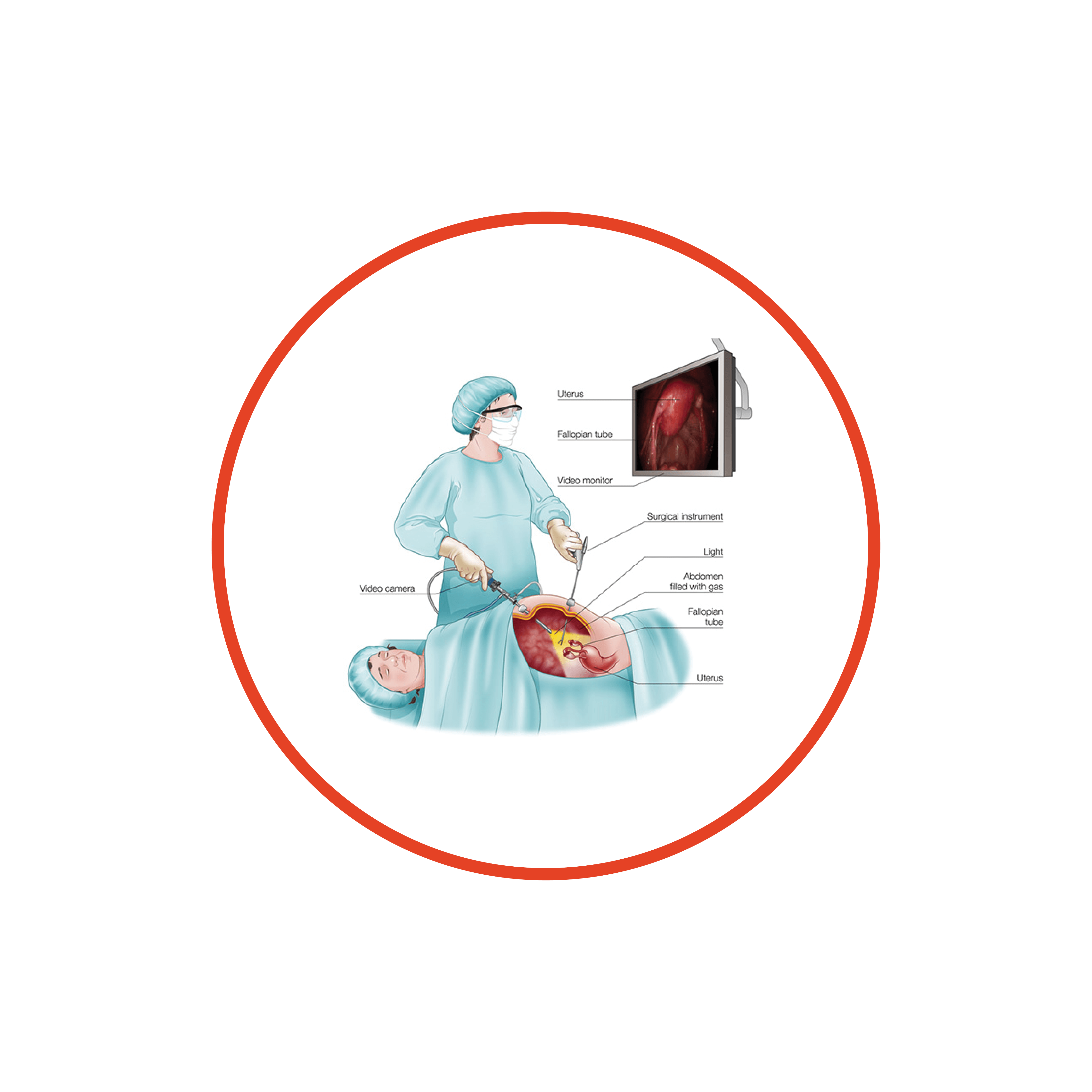
Laparoscopy is commonly called ‘keyhole surgery‘. It is a procedure in which a surgical telescope and video camera is passed through a small cut ‘keyhole‘ in the abdomen, usually in the umbilicus (belly button).
Carbon dioxide gas is used to gently inflate your abdomen during laparoscopy to enable your gynaecologist to see your pelvic organs. This allows your gynaecologist to look at, and operate on, the organs of the pelvis and abdomen. Instruments can be passed through one or more other small cuts in the wall of the abdomen.
The cuts are usually about a centimetre long so the gynaecologist can perform operations without the need for a large cut.
Laparoscopy and keyhole surgical techniques give patients a number of important advantages:
more rapid recovery
reduced pain
smaller scars
Hysterectomy
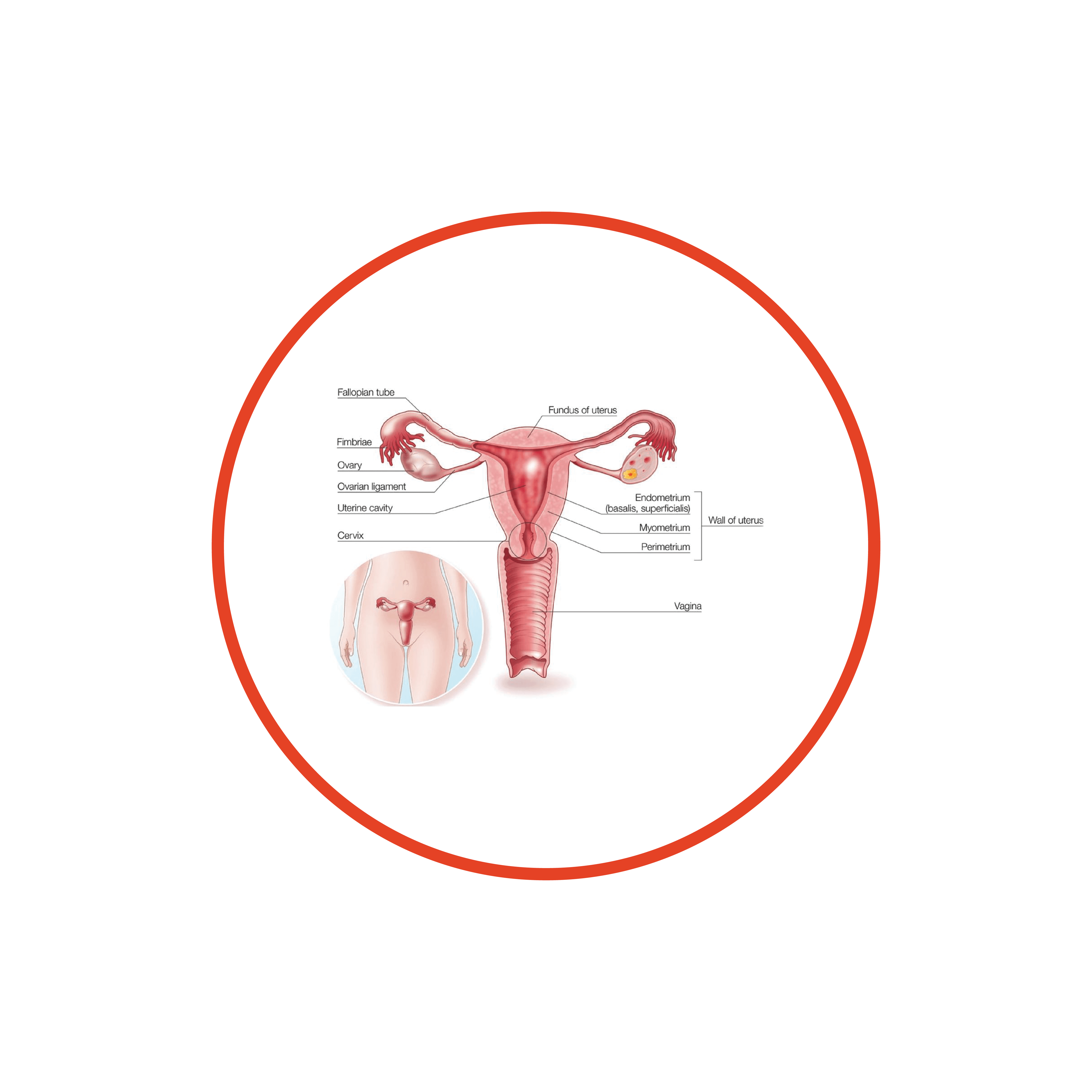
Is an operation where the uterus (womb) is removed.
There are different types of hysterectomy, and during the operation other organs, such as the ovaries or Fallopian tubes – might also be removed.
A total ‘hysterectomy’ means that the uterus and the cervix (neck if the uterus) are removed – this is the most common type of hysterectomy. A ‘subtotal‘ hysterectomy means that the uterus is removed, but the cervix is not – this is a less common operation.
At the time of hysterectomy, one or both of the ovaries might be removed. It is also common for one or both of the Fallopian tubes to be removed. It is important that you are clear about the type of hysterectomy that might be performed, and whether the ovaries or Fallopian tubes are to be removed as well.
vNOTES
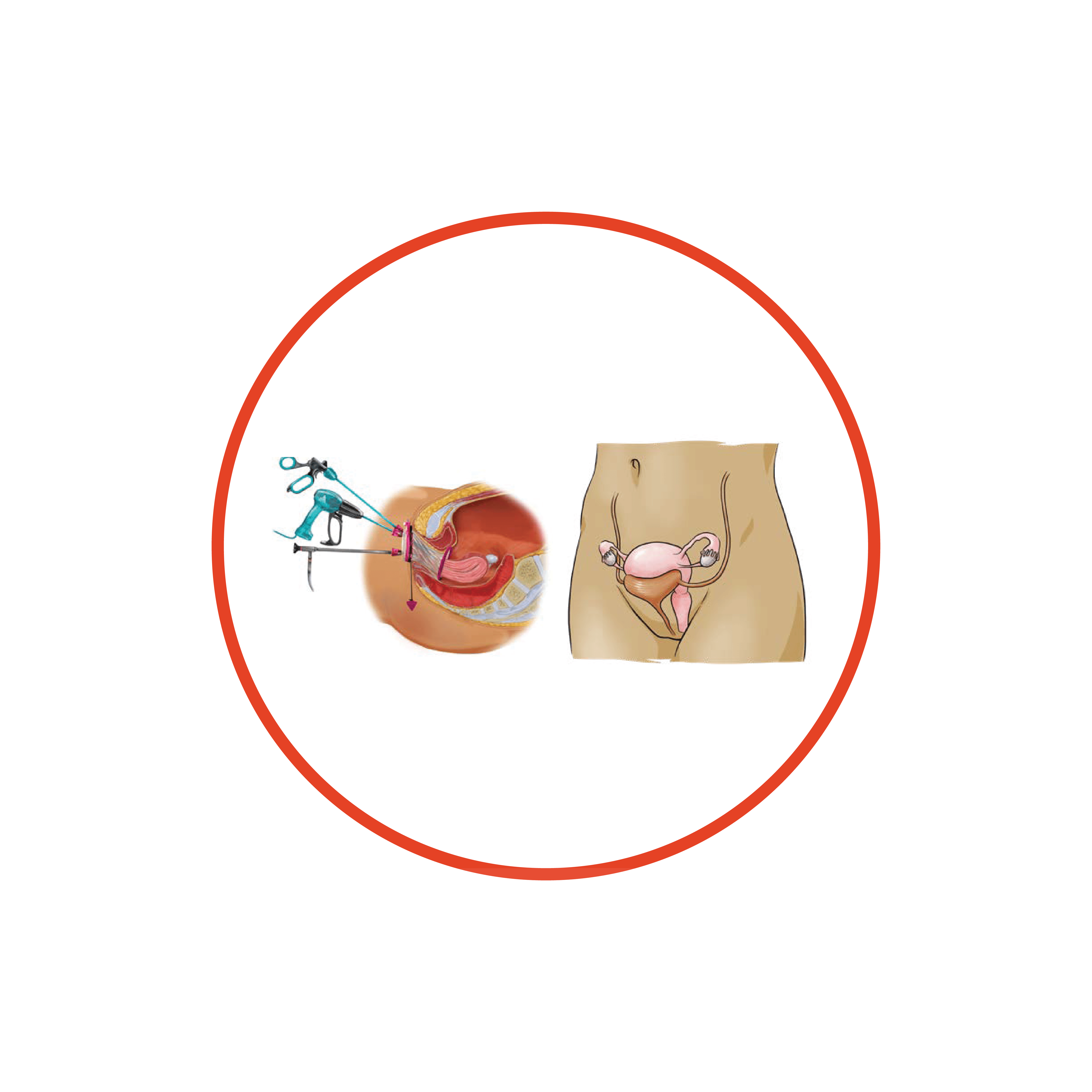
vNOTES (vaginal natural orifice transluminal endoscopic surgery) is a minimally invasive gynecologic approach using the vagina as a surgical access route. Hysterectomy is the most common procedure performed via the vNOTES technique.
vNOTES Procedures include:
Hysterectomy
Salpingectomy
Oophorectomy
Salpingo-oophorectomy
Cystectomy
Conditions treated by vNOTES are:
Adnexal pathology
Abnormal uterine bleeding
Chronic pelvic pain
Fibroids
Prolapse of the uterus
vNOTES has been shown to provide the following benefits to patients compared to the laparoscopic approach:
Shorter hospital stay
Faster recovery time
Less pain medication used
Less postoperative pain
Reduced operating time
Hysteroscopy
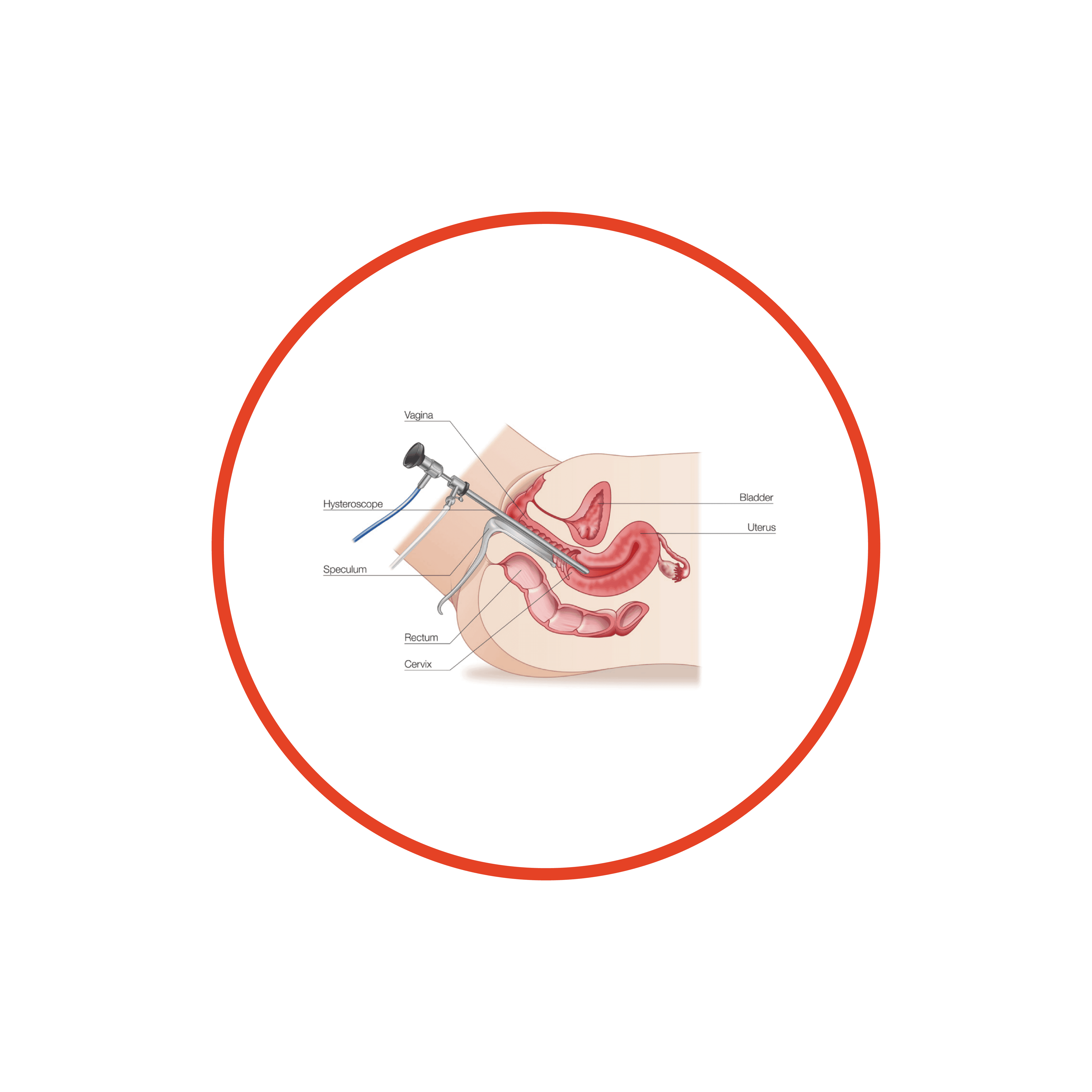
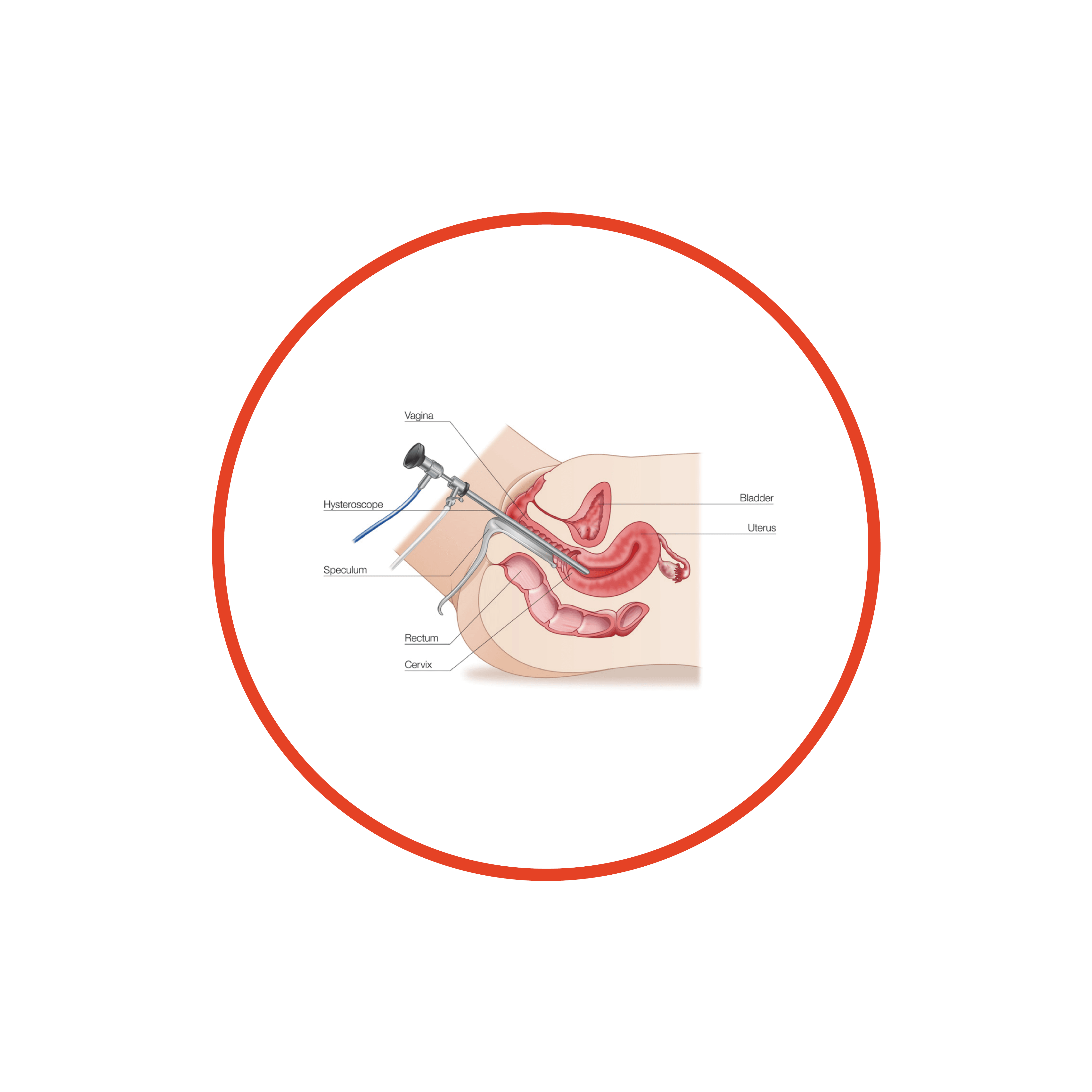
A hysteroscopy is a procedure used to examine the inside of the uterus (womb).
It is carried out using a narrow telescope called the hysteroscopy, which is inserted through the cervix (opening of the womb) into the uterus. The hysteroscope is connected to a light and camera, sending images to a monitor so your gynaecologist can see inside the uterus.
As the hysteroscope is passed into your uterus through the vagina and cervix, no cut needs to be made in your skin.
Common reasons for having a hysteroscopy include abnormal bleeding, fibroids, polyps or difficulty getting pregnant.
Endometrial Ablation
The endometrium is the lining of your uterus (womb). Endometrial ablation is the surgical removal or destruction of this lining. There are different methods of destroying the endometrium including electricity, laser therapy or freezing.
A specialist performs the operation and it is done through the vagina, so there is no need for the abdomen to be cut open. The endometrium will heal leaving scarring, which usually reduces or stops menstrual periods.
In women who have very heavy periods (menorrhagia), an endometrial ablation can be done instead of a hysterectomy as it is an easier procedure than a hysterectomy and is quicker to recover from. Endometrial ablation is only performed in women who no longer wish to have children.
Anterior Vaginal Repair
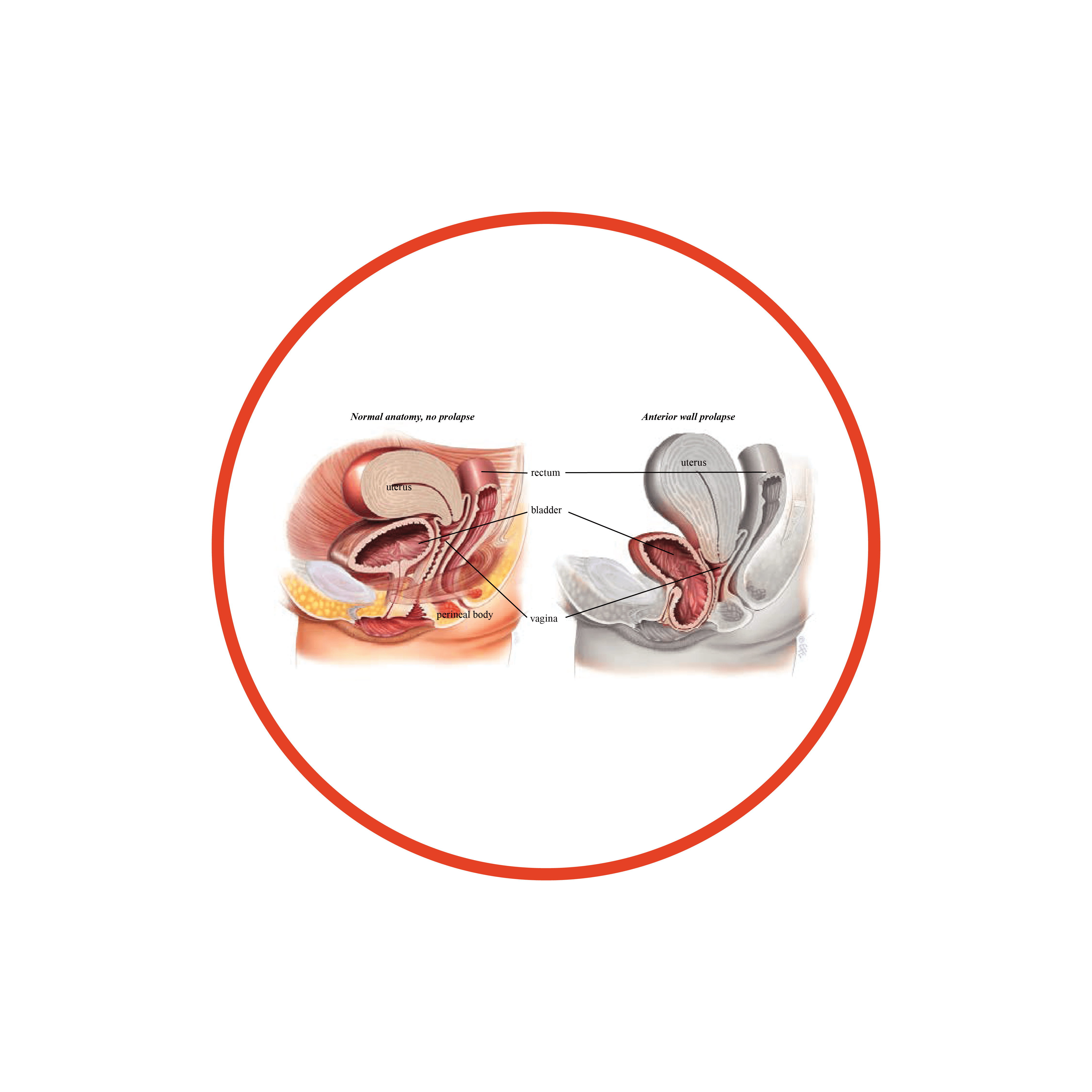
About 1 in 10 women who have had children require surgery for vaginal prolapse. A prolapse of the front (anterior) wall of the vagina is usually due to a weakness in the strong tissue layer (fascia) that divides the vagina from the bladder. This weakness may cause a feeling of fullness or dragging in the vagina or an uncomfortable bulge that extends beyond the vaginal opening. It may also cause difficulty passing urine with a slow or intermittent urine stream or symptoms of urinary urgency or frequency. Another name for an anterior wall prolapse is a cystocele.
An anterior repair, also known as an anterior colporrhaphy, is a surgical procedure to repair or reinforce the fascial support layer between the bladder and the vagina. The aim of surgery is to relieve the symptoms of vaginal bulge and/or laxity and to improve bladder function without compromising sexual function.
Posterior Vaginal Wall and
Perineal Body Repair
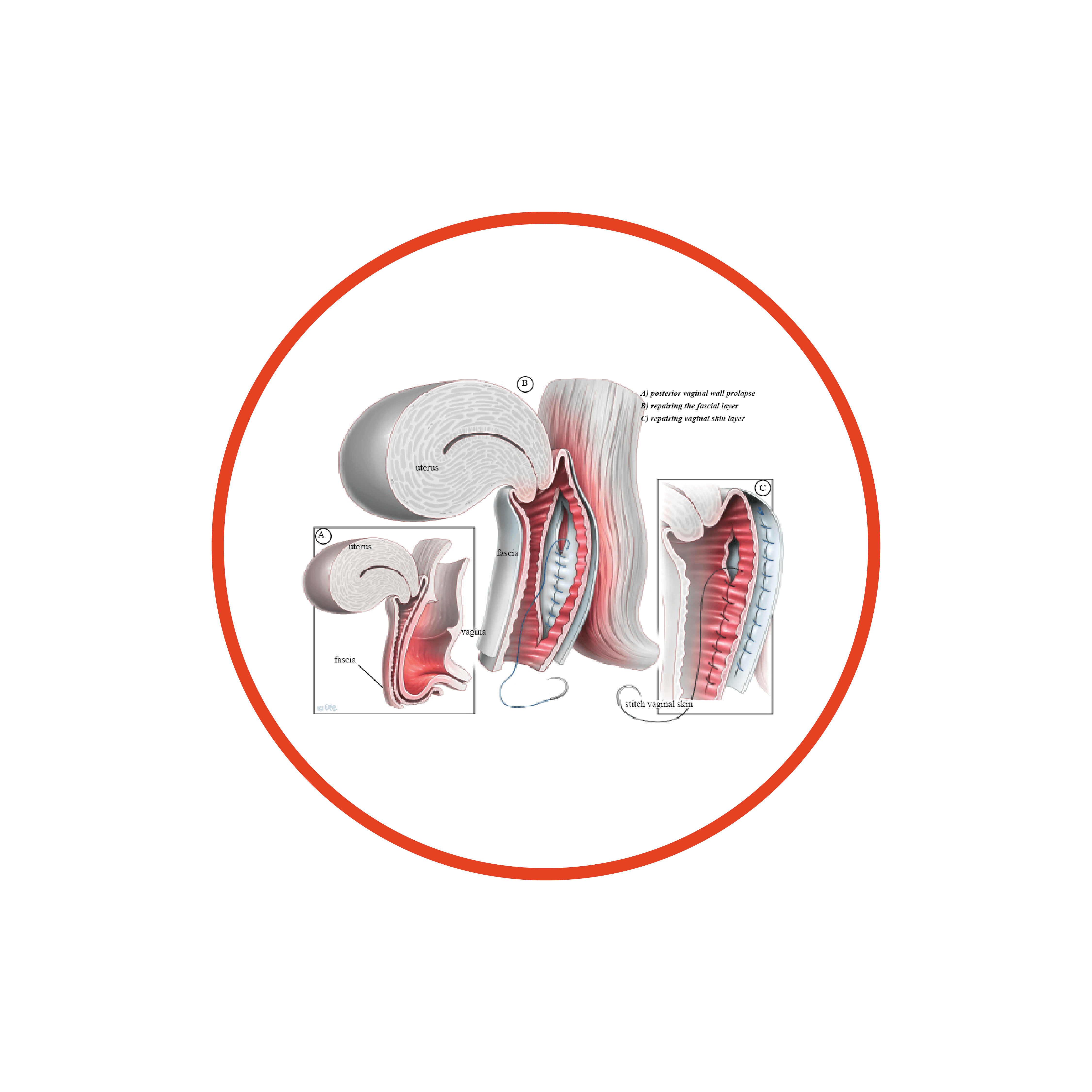
A posterior repair, also known as a posterior colporrhaphy, is a surgical procedure to repair or reinforce the fascial support layer between the rectum and the vagina. A perineorrhaphy is the term used for the operation that repairs the perineal body. The perineal body (the supporting tissue between vaginal and anal openings) also helps to support the back wall of the vagina. The perineum is the area that is often damaged when tears or episiotomies occur during childbirth. This area may need to be repaired along with the back wall of the vagina to give perineal support and in some cases reduce the vaginal opening.
About 1 in 10 women require surgery for vaginal prolapse. A prolapse of the back (posterior) wall of the vagina is usually due to a weakness in the strong tissue layer (fascia) that divides the vagina from the lower part of the bowel (rectum). This weakness may cause difficulty when passing a bowel movement, a feeling of fullness or dragging in the vagina or an uncomfortable bulge that may extend beyond the vaginal opening. Other names for the weakness of the back wall of the vagina include rectocele and enterocele.
Colposcopy
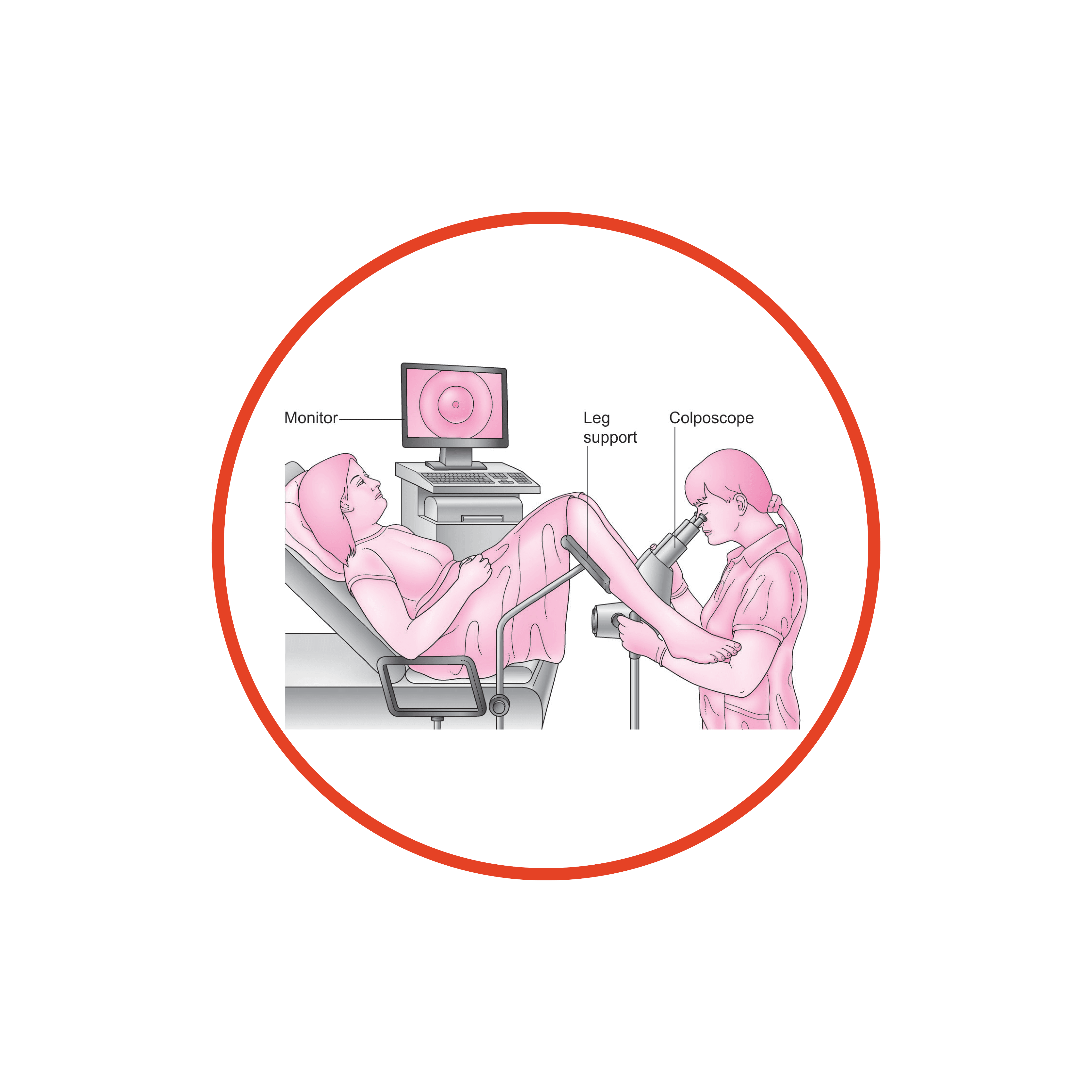
A colposcopy is a detailed examination of the cervix (entrance to the uterus) with a specially lit microscope (colposcope). As with a Pap smear, an instrument called a speculum is inserted into the vagina, and then the colposcope is positioned outside the vagina with its light directed on the cervix.
A specialist will perform a colposcopy if your Pap smear has shown abnormal or cancerous cells on the cervix. During the colposcopy further samples of tissue (biopsies) are usually removed and examined in the laboratory so the doctor can get a clearer idea of the extent of the abnormal cells.
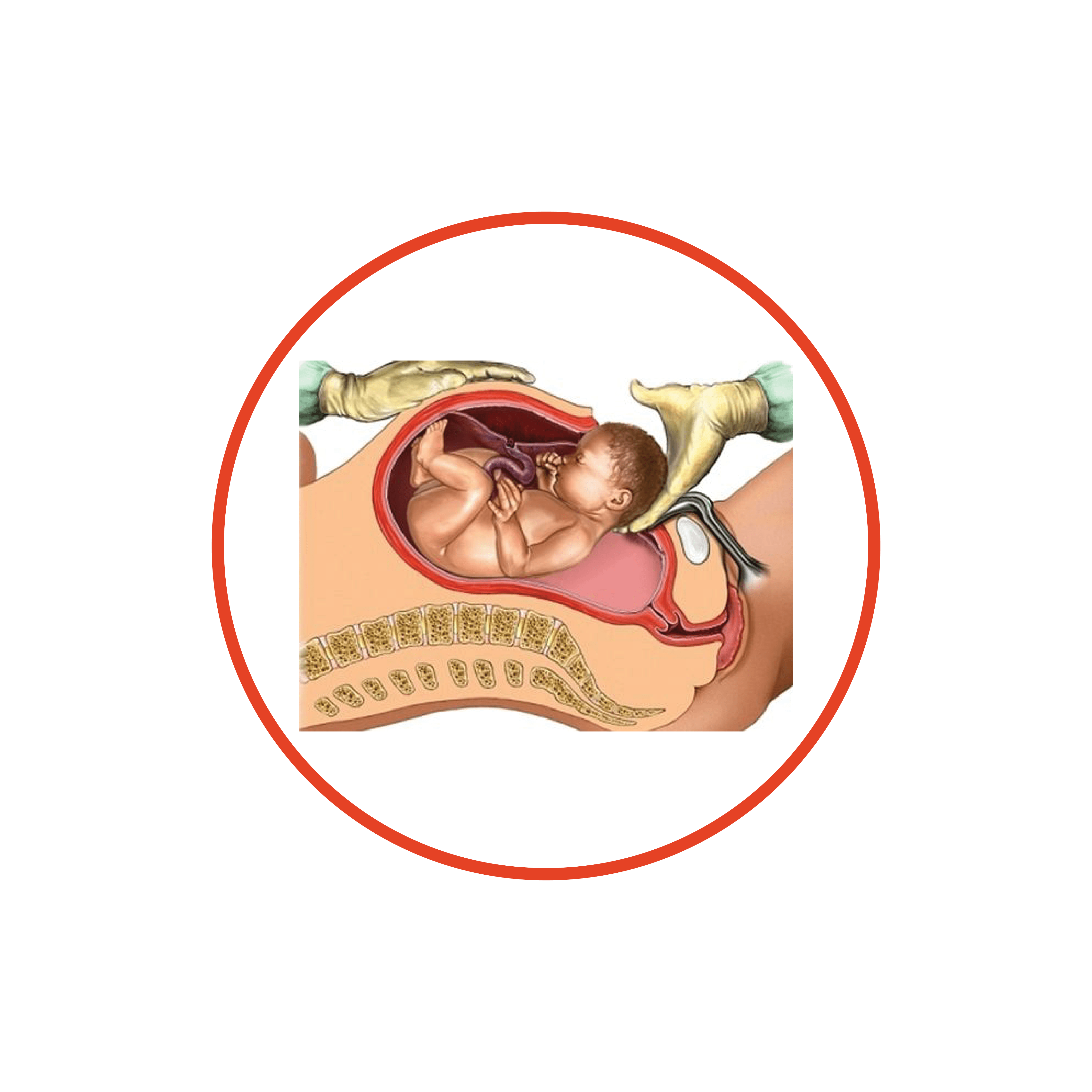
Caesarean Section
A caesarean section is an operation in which a baby is born through an incision (cut) made through the mother’s abdomen and the uterus (womb). The cut is usually made low and around the level of the bikini line.
A caesarean section may be planned (elective) if there is a reason that prevents the baby being born by a normal vaginal birth, or unplanned (emergency) if complications develop and delivery needs to be quick. This may be before or during your labour.
There are several reasons why your obstetrician may recommend an elective caesarean section. Your doctor will discuss the reason for making this decision based on your particular situation and, in some cases, your preferences.
These may include:
you have already had a number of caesarean sections.
your baby is in a breech position (bottom or feet first) and cannot be turned, or a vaginal breech birth is not recommended.
your placenta is partly or completely covering the cervix (opening to the womb).
your baby is lying sideways (transverse) and is not able to be turned by the doctor.
you have a twin pregnancy, with your first baby in a breech position.
CONTACT US
Consult With Us
Dudley Gynaecology is dedicated to a high standard of care in women's health


